New York Cornea Consultants   Aesthetics | Diabetic Retinopathy, Keratoconus and Visian ICL

Pterygium

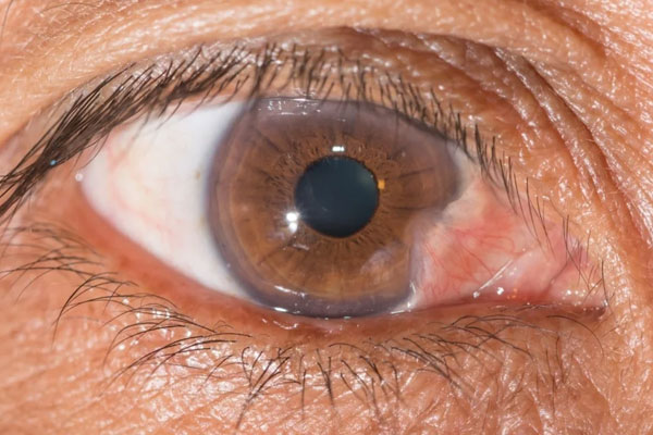
Pterygium (pronounced tur-IJ-ee-um) is an abnormal tissue growth that extends from the corner of the eye toward the center of your eye.

Pterygium

Pterygium (pronounced tur-IJ-ee-um) is an abnormal tissue growth that extends from the corner of the eye toward the center of your eye. The appearance of a pterygium can vary from a small, almost unnoticeable tissue mass to a large, red, noticeable and irritating growth.

Download a pamphlet about AMNIOGRAFT®

Are there ways to prevent it?

Protecting your eyes from prolonged sun exposure is the best prevention method. When outdoors, wear a hat with a brim and UV coated sunglasses to block the sun’s harmful UV rays. These precautions are even more important if you live in a tropical or subtropical area, if you work outside, or if you participate in outdoor activities like fishing, skiing, or gardening.

What are the symptons?

It is possible that a patient with a pterygium has no symptoms at all. Other patients have a variety of symptoms including redness, swelling, itching, irritation, and blurred vision. Vision can be effected if the pterygium grows to cover the center of the eye or if the irregular tissue grows and restricts the eye’s movement.

How is it treated?

Treatment depends on the symptoms. You can treat symptoms with over the counter artificial tears or topical lubricating drops and/or ointments. Your doctor can also prescribe short-term use of topical corticoste anti-inflammatory drops when symptoms are not relieved by over the counter medications. It is also wise to protect your eyes from UV rays. If over the counter and prescription medications do not alleviate your symptoms or if you want a better cosmetic appearance, a pterygium can be surgically removed.

How is a pterygium surgically removed?

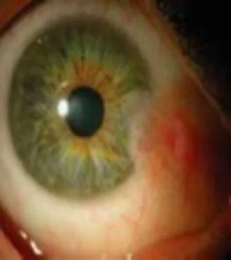
Your pterygium can be surgically removed in an outpatient setting under local or topical anesthesia. Your surgeon will remove all of the pterygium tissue from the surface of your eye. In order to help the area around the pterygium heal, your surgeon will treat the surface of your eye by covering the area where the pterygium was removed with AMNIOGRAFT®.
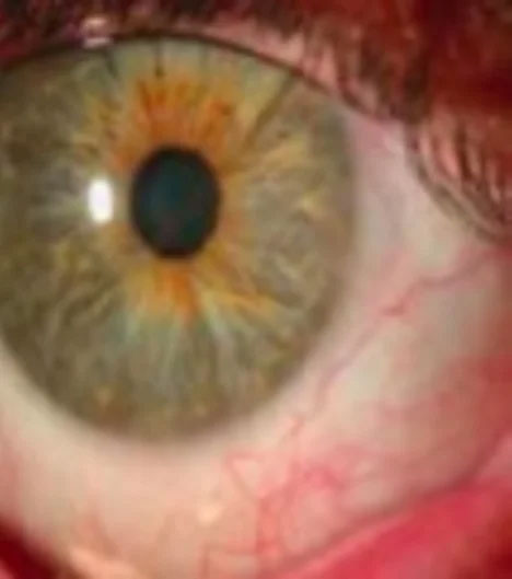
Before AMNIOGRAFT®

New York Cornea Consultants   Aesthetics | Cornea Transplant   Treatment, Keratoconus and Glaucoma

After AMNIOGRAFT®

New York Cornea Consultants   Aesthetics | MIGS Micro-Incisional Glaucoma Surgery, Advanced Dry Eye Treatment and Cataract Surgery

Frequently Asked Questions

What is a pterygium?

+

A pterygium is a benign, wing-shaped growth of conjunctival tissue that extends from the white of the eye toward the cornea. It most often appears on the nasal side of the eye and can range from a small, pale patch to a larger, raised, red lesion that is easily visible. Although benign, the tissue can cause irritation and change the surface of the eye as it progresses.

Pterygia are distinct from pingueculae, which are small, yellow conjunctival deposits that do not invade the cornea. When a pterygium encroaches onto the cornea it can induce astigmatism or interfere with vision by covering the visual axis. Early identification allows monitoring and, if needed, timely intervention to protect vision and comfort.

What causes a pterygium?

+

Chronic ultraviolet (UV) light exposure is the primary risk factor for pterygium formation, which is why the condition is more common in people who spend significant time outdoors. Environmental irritants such as wind, dust, smoke, and dry conditions also contribute by causing chronic conjunctival inflammation. Genetic susceptibility appears to influence who develops a pterygium despite similar exposures.

Age, geographic location in tropical or subtropical climates, and occupations like fishing or farming increase risk due to greater UV and irritant exposure. Repeated minor injury to the ocular surface promotes tissue remodeling that can lead to pterygium growth over time. Protective behaviors and early treatment of surface irritation can reduce the likelihood of progression.

What symptoms should prompt an evaluation for pterygium?

+

You should seek an eye examination if you experience persistent redness, foreign-body sensation, tearing, or chronic eye irritation, especially when symptoms do not respond to lubricating drops. Blurry or distorted vision, especially if you notice changes in how your glasses or contacts correct your vision, warrants prompt assessment. Difficulty moving the eye or a visible fleshy growth approaching the cornea are also reasons to see an ophthalmologist.

Cosmetic concerns are valid reasons for evaluation as well, since an enlarging pterygium can become more noticeable over time. Regular monitoring is important because many patients have minimal symptoms initially but may develop progression that affects vision. An ophthalmologist can document size and growth to guide timing of any needed treatment.

How is a pterygium diagnosed?

+

Diagnosis is primarily clinical and is made during a slit-lamp eye examination that allows detailed inspection of the conjunctiva and cornea. Your doctor will assess the size, vascularity, and extent of corneal involvement and may measure induced astigmatism or changes in the ocular surface topography. Photographs can be taken to document growth over time and support treatment decisions.

If vision is affected, additional testing such as corneal topography, wavefront measurements, or visual acuity and refraction tests may be used to quantify visual distortion. Tear film testing may be performed when dryness is a contributing factor to symptoms. These diagnostic steps help tailor conservative or surgical treatment plans to the individual patient.

What non-surgical treatments are available for pterygium?

+

For many patients, conservative management focuses on symptom relief with preservative-free artificial tears, lubricating ointments, and measures to control ocular surface inflammation. Short courses of topical anti-inflammatory medications, such as corticosteroids, can reduce redness and swelling under a physician's supervision. Managing dry eye and avoiding environmental irritants are important parts of conservative care.

UV protection with wide-brimmed hats and sunglasses that block UVA and UVB rays is a key preventive strategy and complements topical therapies. When irritation persists despite these measures or when a pterygium progresses toward the cornea, the ophthalmologist will discuss surgical options. Regular follow-up allows clinicians to reassess symptoms and intervene at an appropriate time.

When is surgical removal of a pterygium recommended?

+

Surgery is typically recommended when a pterygium causes significant discomfort, chronic inflammation, induced astigmatism, or threatens the visual axis by growing onto the cornea. Limited eye movement due to tissue tightness or recurrent episodes of redness that impair quality of life are also indications. Cosmetic concerns can be a valid reason for elective removal after a thorough discussion with your surgeon.

The decision to operate balances the extent of the lesion, symptoms, the risk of recurrence, and the patient's goals for vision and appearance. An ophthalmologist experienced in anterior segment surgery will review the risks and benefits and recommend the most appropriate timing and technique. Observation remains an option for small, stable pterygia without vision impact.

How is pterygium surgery performed?

+

Pterygium excision is most often done as an outpatient procedure using local or topical anesthesia, providing comfort while the surgeon removes the abnormal conjunctival tissue. After excision, the bare scleral area is commonly covered with a conjunctival autograft taken from the patient's own conjunctiva or with an amniotic membrane graft to promote healing. Fibrin glue or very fine sutures secure the graft and help reduce postoperative inflammation.

Adjunctive measures such as intraoperative mitomycin-C or careful surgical technique are used to lower the risk of recurrence in selected cases. Healing is monitored closely with postoperative eye drops that typically include antibiotics and anti-inflammatory medications to prevent infection and control scarring. Many surgeons use modern grafting methods because they have been associated with lower recurrence and better cosmetic outcomes compared with older techniques.

What are the risks and potential complications of pterygium surgery?

+

As with any ocular surgery, pterygium removal carries risks including infection, bleeding, graft failure, scarring, and discomfort during recovery. Recurrence of the pterygium is the most common complication and can vary depending on surgical technique and individual patient factors. Rarely, surgery can induce significant astigmatism or affect vision if complications occur.

Surgeons minimize risks by selecting an appropriate grafting method, using adjunctive agents when indicated, and providing detailed postoperative care instructions. Patients should follow activity restrictions and medication regimens closely and attend follow-up visits to detect and treat early signs of complications. Open communication with the care team helps ensure issues are addressed promptly and outcomes are optimized.

What should I expect during recovery after pterygium surgery?

+

Recovery typically involves several weeks of reduced activity and the use of prescribed topical antibiotics and anti-inflammatory eye drops to support healing and prevent infection. Patients commonly experience mild discomfort, tearing, light sensitivity, and foreign-body sensation during the first few days to weeks after surgery. Vision may be blurry initially from surface irregularity or medications, with gradual improvement as the eye heals.

Follow-up visits are scheduled frequently in the early postoperative period to monitor graft health, check for signs of recurrence, and adjust treatment as needed. Most people return to nonstrenuous activities within a few days, but strenuous exercise, swimming, and contact lens wear are usually restricted until cleared by the surgeon. Adhering to postoperative instructions and sun protection reduces the chance of complications and supports the best possible visual and cosmetic result.

How does New York Cornea Consultants & Aesthetics approach pterygium care?

+

New York Cornea Consultants & Aesthetics provides individualized evaluation and management of pterygium, combining conservative care with surgical options when indicated to protect vision and ocular surface health. Our team emphasizes detailed diagnostics, personalized treatment plans, and clear explanations so patients understand the rationale for observation versus intervention. We use modern grafting techniques and evidence-based adjuncts to reduce recurrence and promote comfortable, stable healing.

Dr. Michael Ehrenhaus has extensive experience in anterior segment surgery and works with each patient to select the safest, most effective approach tailored to their needs. The practice also stresses preventive education about UV protection and ocular surface maintenance to limit progression and preserve long-term visual quality. Patients are scheduled for regular follow-up to track healing and address concerns promptly.

Schedule a visit today!
Contact Us
SMS Consent
Submit

Thank You!

We appreciate you taking the time to visit our site. We'll review your message and be in touch with you soon.

Continue
New York Cornea Consultants   Aesthetics | Cornea Transplant   Treatment, Cataract Surgery and Advanced Dry Eye Treatment