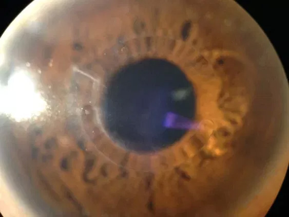
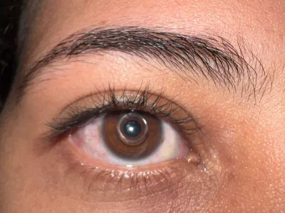
Keratoconus is an eye condition in which the shape of the cornea becomes distorted. The cornea is a clear structure that covers the front of the eye and allows light to enter the eye. In a healthy eye, the cornea curves like a dome. In an eye with keratoconus, the center of the cornea slowly thins and bulges so that it sags and has a cone shape.
Keratoconus tends to affect younger people, and the symptoms sometimes start in the early teen years and progress most rapidly over the next 10 to 20 years. Often, eyeglass prescriptions must be changed frequently as the disease progresses.
Symptoms include:
Difficulty driving at night
Halos and ghosting, especially at night
Eye strain
Headaches and general eye pain
Eye irritation and excessive rubbing of the eye
How is keratoconus diagnosed?
Keratoconus can usually be diagnosed with a slit-lamp examination as well as measurement of the corneal curvature. Your optometrist will look for signs such as corneal thinning, stress lines, and scarring at the apex of the corneal cone. Keratoconus, especially in the early stages, can be difficult to diagnose and its symptoms could be associated with other eye problems. Simply recognizing symptoms does not by itself diagnose the condition.
What is the treatment for keratoconus?
The primary treatment options for keratoconus have traditionally been contact lenses and surgery. In the very early stages of keratoconus, vision problems can be corrected with prescription glasses or contact lenses. As keratoconus progresses, special rigid gas permeable contact lenses may be necessary.
Collagen Cross-Linking is an advanced treatment designed to slow or halt the progression of keratoconus by strengthening the corneal tissue. By combining specially formulated Riboflavin eye drops with carefully controlled ultraviolet light therapy, cross-linking can help stabilize the cornea and preserve vision.
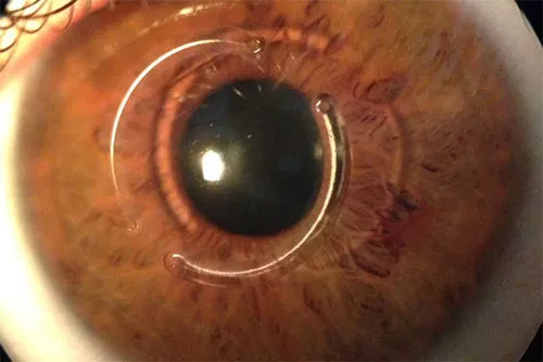
INTACS corneal inserts and Kera-rings are designed to help patients with keratoconus that have become contact lens intolerant. Clinical results have shown both improved lens free as well as lens-wearing vision improvement, and many patients do not necessarily go on to needing a corneal transplant.
Advanced keratoconus, with scarring and thinning of the corneal tissue, may require surgery. Corneal transplant surgery may involve either partial-thickness or full-thickness transplantation depending on the severity of the condition. These procedures are typically performed on an outpatient basis and can significantly improve visual quality and corneal stability.
Please contact the office to make an appointment so that Dr Ehrenhaus can examine you and help advise you with the best options available for your individual needs.
Collagen Cross-Linking (CXL)
Collagen Cross-Linking (CXL) is one of the most important modern treatments for progressive keratoconus. The procedure strengthens weakened corneal tissue and may reduce the likelihood of requiring corneal transplant surgery in the future.
Epi-On Cross-Linking
Epi-on cross-linking preserves the outer surface layer of the cornea during treatment. This minimally invasive technique may provide increased comfort and a faster recovery period for appropriate candidates while helping stabilize corneal progression.
Epi-Off Cross-Linking
Epi-off cross-linking involves gently removing the outer corneal layer prior to treatment to allow deeper penetration of riboflavin into the corneal tissue. This well-established approach is commonly recommended for progressive keratoconus and may provide enhanced corneal strengthening in certain cases.
A comprehensive corneal evaluation helps determine whether epi-on or epi-off collagen cross-linking is the most appropriate treatment approach for each patient.
Introducing CAIRS — An Advanced Treatment Option
In addition to traditional treatments, we now offer Corneal Allogenic Intrastromal Ring Segments (CAIRS) — a cutting-edge procedure for appropriate keratoconus patients who are seeking increased visual stability and comfort.
What is CAIRS?
With CAIRS, ultra-thin ring segments made from donor corneal tissue are placed into a specially prepared channel within your own cornea. These natural implants reshape the corneal curvature and help reduce the bulging and irregular astigmatism characteristic of keratoconus. Because the segments are derived from human tissue rather than synthetic material, there is a potential for greater biocompatibility and long-term stability.
Why Consider CAIRS?
Improved visual clarity: The donor tissue segments can moderate the cone shape and improve how light enters the eye.
Minimally invasive: Compared with full corneal transplant, CAIRS is outpatient and has a quicker recovery period.
Designed for contact-lens intolerant patients: Suitable for those earlier in the disease process who want to preserve more of their own corneal structure.
Added flexibility: CAIRS can be combined with other treatments — such as corneal collagen cross-linking — to both stabilize the cornea and enhance vision.
Is CAIRS Right for You?
We perform a comprehensive evaluation including corneal topography, thickness mapping, and vision testing to determine candidacy. Ideal candidates are patients with progressive ectasia or keratoconus, who may experience contact-lens intolerance or increasing lens changes, and who wish to explore alternatives to more invasive surgery.
Next Step
If you’re managing keratoconus and wish to learn whether CAIRS may be a proper option for you, please schedule a consultation at our office. We’ll review all available treatments and tailor a plan designed specifically to your cornea and visual goals.
Corneal Transplant Surgery
In advanced cases of keratoconus where significant corneal thinning, scarring, or vision loss is present, corneal transplant surgery may become necessary. Corneal transplantation replaces damaged corneal tissue with healthy donor tissue to restore corneal clarity and improve vision.
Depending on the severity and location of the corneal disease, treatment may involve either partial-thickness or full-thickness corneal transplantation techniques. Many corneal transplant procedures are performed on an outpatient basis and can significantly improve visual function and quality of life for patients with advanced keratoconus.
Our practice performs detailed diagnostic testing and comprehensive surgical evaluations to determine the most appropriate treatment plan for each individual patient.
KERARING®
Precise, implantable intrastromal corneal ring segments
KERARING, intrastromal corneal ring segments, offer a minimally invasive approach for corneal remodeling, the treatment of keratoconus, and other ectatic corneal disease. As the most complete and versatile corneal remodeling system available today, KERARING implants are available in variable arc lengths, thicknesses, and diameters. They offer a high level of customization to remodel the corneal surface while reducing refractive errors associated with keratoconus and other ectatic corneal disorders. With implantation, the KERARING segments adjust the cone back in line with the rest of the cornea, regularizing corneal topography and stabilizing corneal ectasia. They offer an effective treatment for managing keratoconus while preventing or delaying the need for more invasive approaches to care.
Quick and Minimally Invasive Procedure
Placing KERARING segments involves a quick and minimally invasive laser procedure, allowing patients to resume their normal activities soon afterward. Typically performed as an outpatient procedure, the resulting topographic and refractive changes start right away, generally stabilizing three months after treatment. It's also reversible if vision changes or the results indicate a need to remove the implants.
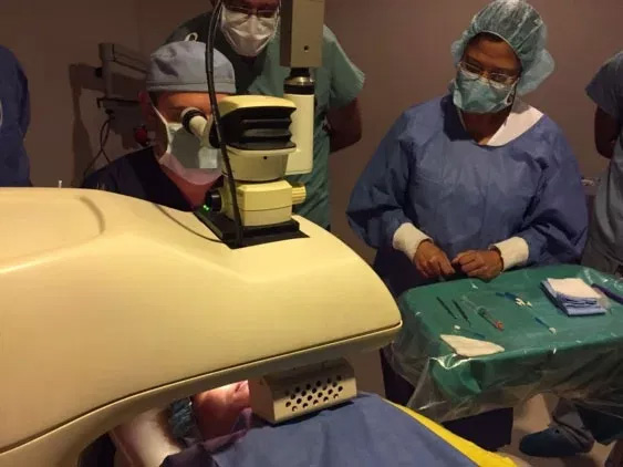
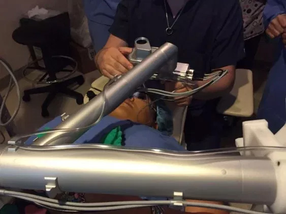
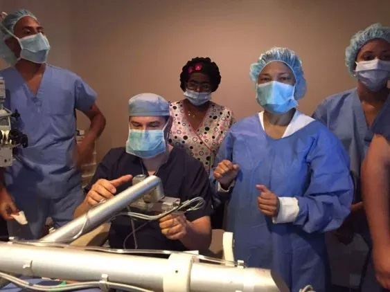
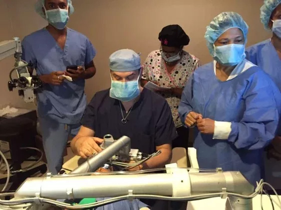
Dr. Ehrenhaus using the latest Femtosecond Laser, designed by Zeimer (Swiss) to implant KERATACx Corneal Rings for Keratoconus and to create bladeless LASIK flaps.
The keratoconus patients implanted with Keratracx corneal ring implants were already noticing up to 2-3 lines of vision improvement without glasses or contacts the next day!
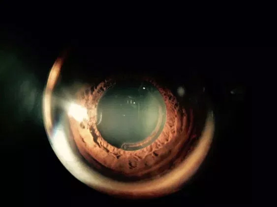
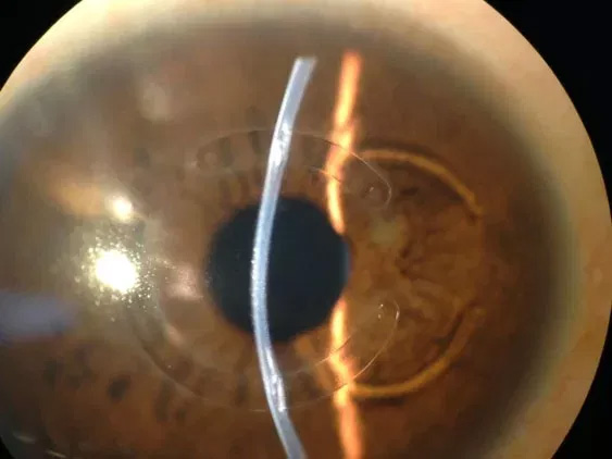
Corneal Rings implanted for Keratoconus
The patient had an easy uncomplicated recovery after the 10 minutes procedure and is already seeing better without his RGP lenses than with them!
LASIK Xtra procedure
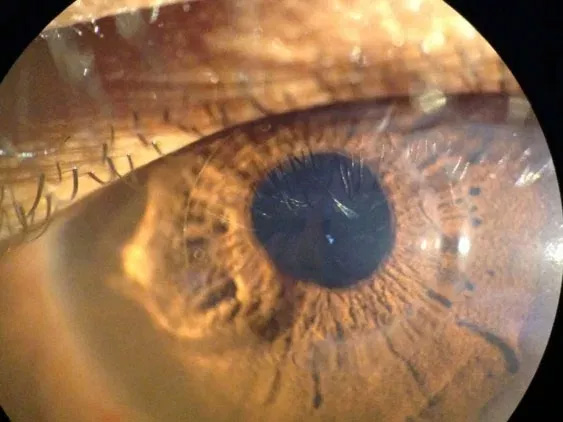
Epithelium-on cornea collagen cross linking for keratoconus
No 9, Calle Republica del Libano Los Jardines Metropolitanos, Santiago
Visit Us Online
Frequently Asked Questions
What is keratoconus?
+
Keratoconus is a progressive condition in which the normally round cornea thins and bulges into a cone-like shape, causing distorted vision. The irregular curvature alters how light is focused onto the retina, producing symptoms such as increased nearsightedness and irregular astigmatism. Changes typically begin in adolescence or early adulthood and can progress over a period of years.
Because the cornea provides much of the eye's focusing power, even small shape changes can have a noticeable impact on vision and visual comfort. The rate of progression varies widely between individuals, so regular monitoring is important. Early detection and appropriate management help preserve vision and minimize the need for more invasive procedures.
What causes keratoconus and who is at risk?
+
The exact cause of keratoconus is unknown, but it appears to result from a combination of genetic, environmental and cellular factors that weaken the corneal structure. A family history of keratoconus increases risk, and the condition is more common in people with certain systemic or ocular conditions, such as atopic disease and eye-rubbing behaviors. Research suggests abnormal collagen and enzymatic activity within the cornea play a role in progressive thinning.
Onset most often occurs during the teen years or early twenties, and progression is usually faster in younger patients. Both males and females can be affected, and severity varies by individual. Identifying risk factors and early signs allows timely intervention to stabilize the cornea and protect vision.
What symptoms should prompt an eye examination for keratoconus?
+
Early symptoms of keratoconus include increasing blurriness, frequent changes in an eyeglass prescription, and difficulty achieving clear vision with spectacles. Patients often report glare, halos, ghosting, or streaking around lights, particularly at night, and may experience eye strain or headaches from visual effort. If symptoms progress quickly or one eye becomes noticeably worse than the other, an appointment with an eye care specialist is recommended.
Because early keratoconus can resemble common refractive errors, subjective symptoms alone do not confirm the diagnosis. A comprehensive exam with corneal imaging helps differentiate keratoconus from other vision problems. Prompt evaluation is important, as interventions such as corneal collagen cross-linking are most effective when applied early in the disease course.
How is keratoconus diagnosed?
+
Diagnosis begins with a comprehensive eye exam that includes refraction and slit-lamp examination to assess corneal shape, thinning and any scarring. Advanced diagnostic tests such as corneal topography, tomography and pachymetry measure corneal curvature and thickness in detail and can detect early or subtle ectatic changes. These imaging tools allow the clinician to map the cornea, monitor progression and plan individualized treatment.
An experienced cornea specialist, such as Dr. Michael Ehrenhaus, will integrate clinical findings, imaging results and the patient’s symptoms to determine diagnosis and stage of disease. Regular follow-up exams with repeat imaging are essential to detect progression and guide timely interventions. Accurate diagnosis also helps determine candidacy for treatments like contact lenses, cross-linking, intrastromal ring segments or transplantation when appropriate.
What nonoperative treatments are available for managing keratoconus?
+
In early or mild keratoconus, vision can often be improved with glasses or soft contact lenses, but as the cornea becomes more irregular, specialty contact lenses may be required. Rigid gas permeable lenses, hybrid lenses and scleral lenses are commonly used to vault or reshape the tear film over the cornea and provide a more regular optical surface for clearer vision. These lens options help many patients achieve functional vision without surgery, though fitting and adaptation may take time.
Corneal collagen cross-linking (CXL) is a minimally invasive procedure that strengthens corneal tissue to slow or halt progression and is frequently combined with optical rehabilitation through contact lenses. In-office fitting by an experienced clinician and ongoing monitoring are important to maintain lens comfort and eye health. Nonoperative strategies focus on stabilizing vision, improving optics and preserving corneal tissue whenever possible.
What surgical and interventional options exist for keratoconus?
+
For patients who cannot achieve adequate vision with contact lenses or who have progressive disease, several interventional options are available to improve corneal shape and visual function. Intrastromal ring segments (such as INTACS or KERARING), femtosecond-laser-assisted ring implantation systems and Corneal Allogenic Intrastromal Ring Segments (CAIRS) can regularize corneal curvature and reduce irregular astigmatism. These procedures are usually outpatient, often reversible, and can delay or prevent the need for full-thickness transplantation.
In advanced cases with significant thinning or scarring, corneal transplantation (full-thickness or lamellar techniques) remains a reliable option to restore corneal clarity and improve vision. Surgical planning balances corneal stability, visual potential and the patient's goals, and may combine treatments—for example, ring segments with subsequent cross-linking—to both reshape and strengthen the cornea. A cornea specialist will discuss expected benefits, recovery and follow-up to tailor the approach to each patient’s needs.
What is corneal collagen cross-linking and who is a candidate?
+
Corneal collagen cross-linking (CXL) is a procedure that uses a photosensitizing agent (riboflavin) and controlled ultraviolet light to strengthen collagen bonds within the cornea, reducing progression of ectasia. The treatment aims to stabilize corneal shape and is most effective when performed early in the disease course or at the first signs of documented progression. Both epithelium-off and epithelium-on protocols exist, and the choice of technique depends on corneal thickness, disease severity and the surgeon’s assessment.
Ideal candidates are patients with documented keratoconus progression or those at risk of deterioration, provided the corneal thickness meets safety thresholds for the selected protocol. Preoperative evaluation includes corneal mapping and thickness measurements to confirm suitability. Following CXL, patients require periodic monitoring to assess stability and to determine whether additional optical rehabilitation is needed.
How does CAIRS differ from synthetic corneal ring segments?
+
Corneal Allogenic Intrastromal Ring Segments (CAIRS) use ultra-thin donor corneal tissue implanted within channels created in the patient’s cornea, whereas traditional intrastromal ring segments are made from synthetic materials. Because CAIRS are derived from human donor tissue, they may offer improved biocompatibility and integration with the host cornea, potentially reducing foreign-body response and improving long-term tolerance. Both approaches aim to reshape the cornea, regularize the surface and reduce irregular astigmatism associated with keratoconus.
Selection between CAIRS and synthetic rings depends on the individual corneal anatomy, degree of ectasia, prior treatments and the surgeon’s experience. CAIRS can be combined with adjunctive procedures such as cross-linking to both stabilize and optimize the corneal shape. A thorough evaluation determines the best option to balance visual improvement, tissue preservation and long-term stability.
Can people with keratoconus maintain good vision long term?
+
Yes. With early detection, appropriate monitoring and timely interventions, many people with keratoconus maintain functional vision for decades. Modern management includes a spectrum of options from specialty contact lenses and cross-linking to ring segments and transplantation, allowing clinicians to customize care based on disease stage and patient goals. The key to long-term success is individualized treatment, adherence to follow-up and addressing risk factors such as chronic eye rubbing.
Outcomes vary among patients, and while some will require more invasive procedures over time, others stabilize with nonoperative measures. Regular visits with a cornea specialist and up-to-date imaging enable proactive adjustments to the treatment plan. Education about the condition and realistic expectations help patients participate actively in their care and optimize visual outcomes.
How should I prepare for a keratoconus evaluation and what does follow-up care involve?
+
To prepare for a keratoconus evaluation, bring a current list of eyewear, contact lens history and any prior corneal imaging if available, and avoid wearing rigid contact lenses for the recommended time before testing to ensure accurate measurements. The evaluation typically includes visual acuity testing, refraction, slit-lamp exam and corneal topography/tomography and pachymetry, which together assess shape, thickness and signs of progression. The clinician will review results, discuss potential treatments and outline a personalized monitoring schedule.
Follow-up care depends on disease activity and chosen therapy, ranging from periodic imaging every few months to annual checks for stable cases. After procedures such as cross-linking or ring implantation, more frequent early visits assess healing, visual changes and the need for optical rehabilitation. The office of New York Cornea Consultants & Aesthetics emphasizes clear communication and coordinated follow-up to ensure optimal long-term care for patients with keratoconus.