Dr. Ehrenhaus will be happy to sit and discuss with you the best options for your individual case and the surgery most likely to help you have a more rapid recovery.
Whether you are a candidate for the new technique of sutureless corneal transplantation, or a candidate for a lamellar transplant designed for your eyes with the femtosecond IntraLase laser, or you are a candidate for a full thickness corneal transplant, please feel free to contact us to discuss your case, your history and your concerns.
Dr. Ehrenhaus will be happy to sit and discuss with you the best options for your individual case and the surgery most likely to help you have a more rapid recovery.

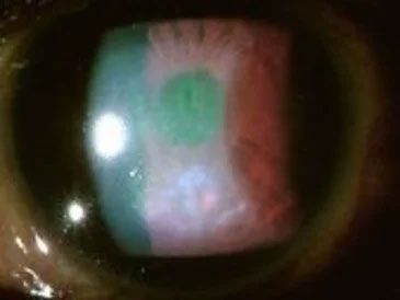

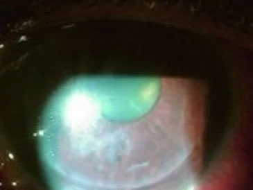

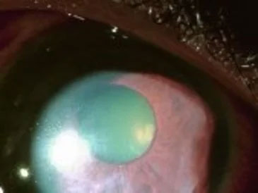
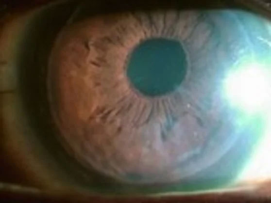
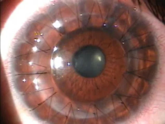

A cornea transplant replaces damaged or diseased corneal tissue with healthy donor tissue to restore clarity and improve vision. This procedure is recommended when the cornea becomes scarred, warped, or dysfunctional from conditions such as advanced keratoconus, endothelial disease, trauma, or infection that do not respond to medical therapies.
The decision to proceed with transplantation is individualized and based on visual function, symptoms, and the health of the rest of the eye. Your surgeon will review diagnostic testing, prior treatments, and overall ocular health to determine whether a transplant offers the best chance for visual rehabilitation.
There are several transplant techniques tailored to the location and depth of corneal damage, including full-thickness penetrating keratoplasty (PKP) and partial-thickness or lamellar procedures such as DSAEK/DSAEK and DMEK. Lamellar techniques replace only the diseased layers of the cornea and often provide faster recovery and reduced risk compared with full-thickness grafts.
Advances such as femtosecond laser–assisted lamellar cuts and sutureless transplantation have expanded options for precise tissue preparation and improved wound stability. Your surgeon will recommend the procedure that best matches the pattern of damage, expected outcomes, and your individual needs.
Sutureless corneal transplantation techniques, commonly used with modern endothelial grafts like DSAEK and DMEK, secure the donor tissue in place without the long-term presence of surface sutures. These methods typically rely on an internal tamponade (for example, an air or gas bubble) and careful postoperative positioning to allow the donor layer to adhere to the host cornea.
Because surface sutures are minimized or avoided, patients often experience less surface irritation and a more rapid visual rehabilitation compared with traditional sutured PKP. However, the choice between sutureless and sutured approaches depends on the type of disease, graft layer required, and surgeon assessment of the best trade-offs for each case.
Candidacy is based on a thorough clinical examination and diagnostic testing including corneal topography, pachymetry, endothelial cell counts, and slit-lamp biomicroscopy to define which corneal layers are affected. Lamellar procedures are preferred when disease is limited to the anterior or posterior layers, while full-thickness PKP is reserved for cases with widespread scarring or structural compromise of multiple corneal layers.
At New York Cornea Consultants & Aesthetics, Dr. Michael Ehrenhaus discusses the risks, benefits, and expected recovery for each option and tailors recommendations to the patient’s medical history and visual goals. A collaborative discussion also includes how other ocular conditions could affect outcomes and whether combined procedures are advisable.
The preoperative evaluation includes a detailed medical and ocular history, a comprehensive eye exam, and specialized testing such as corneal imaging, endothelial cell analysis, and measurements of corneal thickness and curvature. Your surgeon will review medications, systemic health issues that may affect healing, and prior eye surgeries to plan the safest and most effective approach.
Patients are also counseled about the postoperative course, positioning requirements for procedures that use an internal air or gas bubble, and the follow-up schedule needed to monitor graft adherence and ocular pressure. Clear preoperative communication helps set realistic expectations and optimizes readiness for surgery.
DSAEK (Descemet stripping automated endothelial keratoplasty) selectively replaces the diseased endothelial layer and a thin portion of posterior stroma, whereas penetrating keratoplasty replaces the full thickness of the cornea. Because DSAEK is tissue-sparing and often performed through smaller incisions, it typically produces faster visual recovery and reduced surface-related complications compared with full-thickness grafts.
Recovery after DSAEK often involves a shorter period of visual fluctuation and fewer suture-related visits, but patients still require close monitoring for graft adherence, rejection, and intraocular pressure changes. The choice between DSAEK and PKP depends on the underlying condition, corneal anatomy, and surgeon judgment.
Management strategies include topical and, when indicated, systemic anti-inflammatory therapy, close surveillance with frequent follow-up visits, and timely surgical intervention when needed. Your surgeon will outline warning signs to report immediately and a follow-up plan to identify and treat complications early.
Early recovery varies by procedure: lamellar procedures such as DSAEK often show visual improvement within days to weeks, while full-thickness PKP may take several months to achieve a stable visual result. Initial postoperative visits focus on assessing graft attachment, controlling inflammation, and monitoring intraocular pressure to ensure a healthy healing environment.
Suture removal, if applicable, and optimization of vision with glasses or contact lenses may occur over several months to a year depending on healing and astigmatism. Regular follow-up is essential during this period to detect rejection, adjust medications, and plan any rehabilitative measures to maximize visual outcomes.
Some early or mild corneal conditions can be managed with non-surgical options such as specialty contact lenses, corneal collagen cross-linking for progressive keratoconus, medication for ocular surface disease, and intracorneal ring segments for certain corneal shape irregularities. These measures can delay or, in some cases, avoid the need for transplantation by strengthening the cornea or improving surface health.
When conservative measures are insufficient, minimally invasive procedures and modern lamellar transplants offer targeted surgical options with favorable recovery profiles. A thorough evaluation helps determine whether non-surgical therapies are appropriate or whether surgical referral for transplant is the best path to restore vision.
Donor tissue is obtained from accredited eye banks that screen for systemic and ocular diseases and assess tissue quality, including endothelial cell density and corneal integrity. Eye banks follow strict protocols for tissue recovery, testing, preservation, and documentation to maximize safety and suitability for transplantation.
Surgeons work with eye banks to select graft tissue that best matches the needs of the recipient procedure, whether that requires high endothelial counts for endothelial grafts or full-thickness clarity for PKP. Proper tissue handling and timely transplantation are critical to graft viability and long-term outcomes.
Dr. Michael Ehrenhaus and the clinical team emphasize individualized care, taking time to explain the condition, available treatment options, and expected recovery so patients can make informed decisions. The practice coordinates preoperative testing, perioperative instructions, and a structured follow-up schedule to monitor healing and manage any complications promptly.
Postoperative support includes timely clinic visits, clear medication plans, and education on warning signs that require urgent attention. By combining advanced surgical techniques with attentive follow-up, the team aims to optimize visual outcomes and overall eye health for each patient.